NEUWAVE-12
Welcome to NEUWAVE-12!

The 12th edition of the Neutron Wavelength-dependent Imaging Workshop (NEUWAVE-12) is set to take place from September 1st to September 4th, 2024, in Lund, Sweden.
The NEUWAVE workshops gather prominent researchers worldwide in the field of energy-resolved neutron imaging, a cutting-edge methodology leveraging the energy/wavelength dependency of neutron transmission for enhanced quantitativity. NEUWAVE strives to promote collaboration and advance energy-resolved neutron imaging by facilitating the exchange of information and experiences in an intimate workshop setting. Key topics of the NEUWAVE workshop encompass the current status of neutron imaging instruments, the evolution of imaging methods (such as Bragg-edge imaging, resonance absorption imaging, and imaging utilizing neutron interference effects or polarized neutrons), the advancement of detectors and instrumentation, and applications of energy-resolved techniques.
The ODIN instrument at the European Spallation Source (ESS), set to receive first neutrons next year, can be considered as one of the outcomes of the discussions and collaborations nurtured by NEUWAVE, exemplifying the success of the NEUWAVE workshop series.
Previous NEUWAVE workshops were held in various locations:
- 2008: Garching, Germany
- 2009: Abingdon, UK
- 2010: Sapporo, Japan
- 2011: Gatlinburg, USA
- 2013: Lund, Sweden
- 2014: Garching, Germany
- 2015: Mito, Japan
- 2016: Abingdon, UK
- 2017: NIST, USA
- 2019: PSI, Switzerland
- 2023: Tokyo, Japan
NEUWAVE-12 will commence with the traditional walking discussion on Sunday, September 1st. We will take the ferry to the beautiful island Ven, formerly home to danish astronomer Tycho Brahe.
The main workshop sessions are scheduled from September 2nd to 4th, featuring both oral and poster presentations, as well as open discussions. Following the NEUWAVE tradition, a limited number of oral presentations have been accepted to allow substantial discussion time for each topic.
The first two days of the main workshop, September 2nd and 3rd, will take place at AF Borgen in central Lund, while the last day, September 4th, is scheduled to take place at ESS and will include a tour of the facility (a unique chance before the start of operations!). A workshop dinner, sponsored by Amsterdam Scientific Instruments, is planned for September 3rd at Hos Talevski i Stadsparken.
In addition to the main workshop, a satellite workshop on NCrystal (library for thermal neutron transport in crystals and other materials) will be held at LINXS (Lund Institute of Advanced Neutron and X-ray Science) on September 5th.
NEUWAVE-12 is organized and sponsored by the European Spallation Source. Additional support is provided by Amsterdam Scientific Instruments and LoskoVision, who will respectively sponsor the dinner and a prize for the best poster.


-
-
10:00
→
20:00
Walking Discussion at Ven
-
10:00
→
20:00
-
-
08:30
→
09:00
Registration and Poster setup
-
09:00
→
09:50
OpeningConvener: Robin Woracek (European Spallation Source ERIC)
-
09:00
Welcome and Logistics 10mSpeaker: Robin Woracek (European Spallation Source ERIC)
-
09:10
Welcome to ESS and its future Science 20mSpeaker: Giovanna Fragneto (European Spallation Source ERIC)
-
09:30
Poster Pitches 20m
-
09:00
-
09:50
→
11:00
Morning Session 1Convener: Burkhard Schillinger (Heinz Maier-Leibnitz-Center (FRM II), Technische Universität MNünchen, Germany)
-
09:50
Progress in wavelength resolved imaging at PSI 25m
This presentation will give an overview of wavelength resolved imaging methods and instru-mentation developed and pursued at the instruments of the Applied Materials Group at PSI. It will focus on such methods, techniques, applications and results not covered in other con-tributions. Recently added capabilities from a new velocity selector, and new double crystal monochromator at ICON as well as the addition of a chopper system for our pioneering ap-proaches for time-of-flight imaging at a continuous source will be discussed. New contrast modalities such as inelastic scattering contrast, polarized dark-field imaging as well as multi-directional dark-field contrast will be presented together with outstanding applications in material science[1].
Speaker: Markus Strobl -
10:15
Pulsed neutron imaging activities at RADEN in J-PARC 25m
The Energy-resolved neutron imaging system, RADEN [1], has been constructed in J-PARC MLF in 2014 and started the user operation in 2015, and we will soon celebrate the 10th year from the successful first neutron beam extraction in RADEN. So far, we continued developments on energy-resolved neutron imaging techniques using time-of-flight (TOF) analysis and on the devices related to neutron imaging. Accordingly, now RADEN becomes a unique instrument that can cover Bragg edge, resonance absorption, polarimetry, and grating interferometry imaging techniques with wavelength/energy resolution. Regarding the application, more than half of proposals submitted to RADEN use TOF technique, especially Bragg edge imag-ing, and remaining proposals are mostly special neutron radiography/tomography experi-ments, such as high-resolution imaging and in-situ/in-operando imaging, which are difficult to conduct in other neutron imaging facilities in Japan. Especially, this situation becomes more obvious after the successful operation restart of the Japanese research reactor, JRR-3, in 2021, which was suspended more than 10 years due to fitting the new regulatory standard created after the Great East Japan Earthquake in 2011. Thus, the major neutron imaging facili-ties in Japan are all in operation, and hence, cooperation and distinction among facilities be-came even more pronounced, and by taking advantage of their features, it allows us their ef-ficient use in a wide range of experiments.
Recently a new neutron imaging instrument in MLF was proposed, which views the coupled hydrogen moderator of JSNS to obtain higher neutron flux with moderate wavelength resolu-tion, to compensate RADEN. In addition, because Kyoto University Research Reactor (KUR) is scheduled to be shut down in May 2026 due to the deadline for returning spent fuel to the United States, the Japanese government authorized a project to construct a new research re-actor at the site of the fast breeder reactor ”Monju” in Fukui prefecture to be a base of neu-tron science in western Japan on behalf [2]. We are proposing to install two neutron imaging instruments in this new research reactor each of thermal and cold neutron beams.
In the presentation, we will discuss the current status of RADEN and what we learned through the 10-year operation, in addition, our future prospects on RADEN and Japanese neutron im-aging activity.References
[1] T. Shinohara, et al., Rev. Sci. Instrum. 91, 043302 (2020)
[2] https://www.jaea.go.jp/04/nrr/en/Speaker: Takenao Shinohara (Japan Atomic Energy Agency) -
10:40
Poster Pitches 20m
-
09:50
-
11:00
→
11:40
Coffee Break & Posters 1 40m
-
11:40
→
12:30
Morning Session 2Convener: Nikolay Kardjilov (Helmholtz-Zentrum-Berlin (HZB))
-
11:40
Commissioning the VENUS beamline: preliminary results 25m
The VENUS hyperspectral imaging beamline is currently being commissioned at the Oak Ridge National Laboratory Spallation Neutron Source after several years of construction. The beamline provides collimation ratios varying from 400 to 2000. It is equipped with choppers, a cadmium filter, and two collimators to reduce background in the instrument cave. While the flight tubes located in the front-end are evacuated, the cave flight tubes are filled with helium to minimize the thickness of the aluminum window near the sample area. VENUS is optimized for Bragg edge and resonance imaging, with a 20 x 20 and 4 x 4 cm2 field-of-view, respectively. VENUS is equipped with three detectors: the ANDOR charge-coupled device (CCD) iKon-XL 230, the QHY scientific Complementary Metal Oxide Semiconductor (sCMOS) sensor 6060, the microchannel plate Timepix detector (with a maximum field-of-view of 2.8 x 2.8 cm2). A microchannel plate Timepix 3 detector is also being commissioned. Figure 1 displays the general layout of the beamline with the front-end area (where optical components, the filter, and choppers are located), the cave and beam stop, the radiological materials area (RMA), and the control hutch. During routine operations, the cave, RMA and control hutch are accessible.
The instrument commissioning focuses on the performance of the beamline components, the study of the moderator, the acquisition and data workflows, calibration measurements, and early scientific results. Some of these preliminary results are presented here.Speaker: Jean Bilheux (Oak Ridge National Laboratory) -
12:05
Recent advances in neutron imaging at FRM II 25m
The neutron imaging group at FRM II, Garching operates the two neutron imaging facilities ANTARES and NECTAR. ANTARES provides a cold neutron spectrum, which gives high sensitivity for even small changes of composition in a sample and is used for neutron imaging with high spatial resolution as well as advanced techniques such as imaging with polarized neutrons or neutron grating interferometry (nGI). The instrument NECTAR is a unique facility that provides a fast fission neutron spectrum that allows the investigation of even very bulky samples and shows contrast complementary to X-rays or gammas. Additionally, thermal neutrons and gamma radiography are also available at NECTAR in a multi-modal approach.
While FRM II has not been running due to repair works for an extended period of time, we have performed many upgrades to our instruments and have performed many experiments at other facilities to support ongoing internal and user projects such as the development of event-mode neutron imaging detectors, studying the defect evolution in additively manufactured samples, investigating the process of freeze drying with neutron imaging or using neutron grating interferometry to track the magnetic domain behavior in electric steel under tensile stress. Moreover, we have proposed to install the additional and complementary neutron imaging instrument FLASH-NT on a cold neutron guide end position at FRM II in the framework of an upgrade program.
In our contribution, we will give an overview of recent achievements and activities of the neutron imaging group at FRM II and show the new experimental possibilities users will have at our instruments.Speaker: Michael Schulz (Heinz Maier-Leibnitz Zentrum (MLZ), Technische Universität München (TUM))
-
11:40
-
12:30
→
12:40
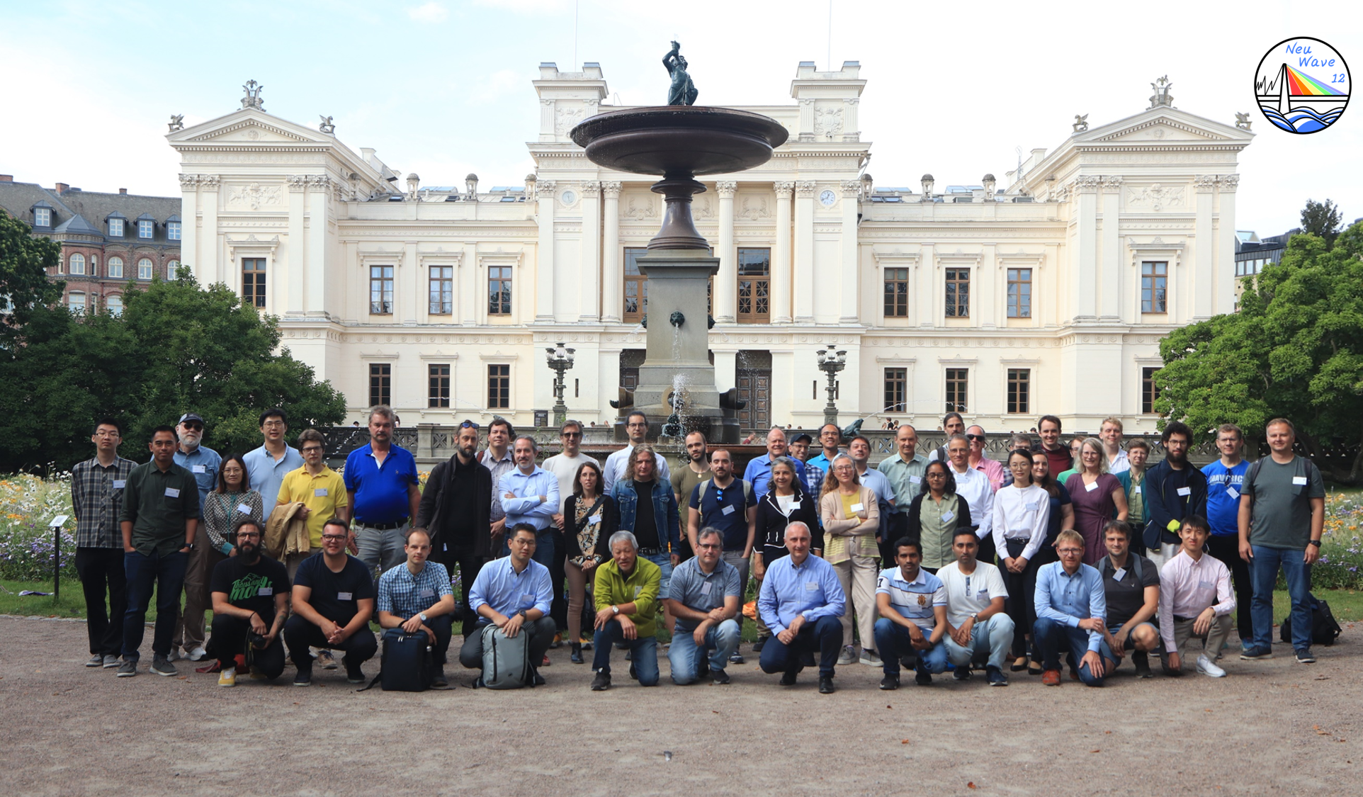
Group Photo!! 10m
-
12:40
→
14:30
Lunch in Lund restaurants 1h 50m
-
14:30
→
16:10
Afternoon Session 1Convener: Takenao Shinohara (Japan Atomic Energy Agency)
-
14:30
Non-destructive reconstruction of bulk microstructure and interlayer diffusion in wire‑arc additive manufactured materials 25m
Energy-resolved neutron imaging provides unique opportunity to investigate bulk microstructure and elemental composition, all in one non-destructive measurement, providing neutron transmis-sion spectra can be measured in a wide energy range [1]. The existence of bright pulsed neutron beams and fast neutron counting detectors enable reconstruction of sample characteristics within several centimetre areas with ~0.1 mm resolution measured all at the same time.
We present the results of experiments where Al alloy samples were produced by wire-arc additive manufacturing (AM) technique [2]. Interdiffusion of materials between the deposition layers was investigated through neutron resonance absorption, while the microstructure was revealed through Bragg edge imaging. Several samples were used in this study, some with rolling applied to individual layers during manufacturing process. Both as-built and heat treated samples were investigated. The analysis of neutron resonance absorption enabled quantitative reconstruction of Ag and Cu elemental composition within different layers of AM printed materials.Speaker: Anton Tremsin (University of California at Berkeley) -
14:55
Study on deformation twinning in Magnesium and its alloy: Combined neutron Bragg-edge imaging and diffraction 25m
Neutron Bragg-edge imaging, offering high spatial resolution for visualizing crystallographic information, has become a useful tool for material research. Magnesium (Mg) and its alloy, as one of the light structural materials, have been widely applied in various industries. Deformation twinning plays an important role in the deformation processes of Mg alloys with a hexagonal-close-packed (HCP) structure. Our study focuses on understanding the mecha-nisms of deformation twinning in Mg and its alloy using neutron Bragg-edge imaging and diffraction techniques. Two types of samples were designed: Pure Mg with coarse grains, for direct observation of deformation twins using energy/wavelength-dependent neutron trans-mission imaging; and Mg-6Zn alloy with fine grains, for extracting the volume fraction of de-formation twins via Bragg-edge transmission spectra analysis. Rectangular-shaped pure Mg and Mg-6Zn (6 wt.% Zn) alloy samples were prepared. Compression tests were carried out using a loading machine with different strains. Ex-situ pulsed neutron transmission imaging experiments were conducted at BL22 RADEN of the MLF/J-PARC [1]. A µNID detector was used to obtain 2D neutron Bragg-edge spectra of the samples after compression [2], and GUI-RITS software was used for Bragg-edge spectral analysis [3].
As shown in Fig. 1, the transmission spectra of the Mg-6Zn sample along the compression direction exhibit evident changes with increasing strain, especially for the three prominent Bragg edges. The height of the {0002} edge increases with strain, while the {10-10} edge de-creases simultaneously, indicating texture evolution due to twinning. The volume fraction changes of {10-12} <10-11> twins were quantitatively assessed through Bragg-edge spectral analysis. Additionally, direct visualization of twin formation and growth during compression of coarse-grained pure Mg was achieved using wavelength-dependent imaging. Comparative analyses with in-situ neutron diffraction data from BL19 TAKUMI, MLF/J-PARC, will also be presented.
References
[1] T. Shinohara et al., Rev. Sci. Instrum. 91, 043302 (2020)
[2] J.D. Parker et al., JPS Conf. Proc. 22, 011022 (2018)
[3] K. Oikawa et al., J. Phys. Conf. Ser. 2605, 012013 (2023)Speaker: Yuhua Su (Japan Atomic Energy Agency) -
15:20
Strain and texture mapping using Frame Overlap Bragg Edge Imaging (FOBI) at SINQ 25m
Recent advances have been made at SINQ, the neutron source of the Paul Scherrer Institute in Villigen, Switzerland, in applying the Frame Overlap Bragg edge Imaging (FOBI) method for non-destructive internal strain measurements [1]. This method employs a multiple slit chopper with a pseudorandom pattern, allowing neutron pulses with different wavelengths to arrive at the detector simultaneously. The overlapping spectra prevent direct conversion to wavelength, so a coded source imaging technique is used to disentangle the overlap be-tween different pulses [1]. This approach compensates for flux reduction by increasing the duty cycle, thereby reducing measurement times. FOBI has been successfully employed at POLDI (Pulse OverLap DIffractometer), the Time-Of-Flight (TOF) diffractometer at SINQ, for texture determination and strain mapping in various metallic alloys produced by additive manufacturing.
In this work, we present the application of strain and texture mapping using the FOBI meth-od at POLDI in multi-material lattice structures produced by laser powder bed fusion. Addi-tionally, we study the internal strain during in-situ lap shear tests on different combinations of dissimilar materials joined by friction stir welding.
Speaker: Sergio Soria (Applied Materials Group, Paul Scherrer Institut, Villigen, Switzerland) -
15:45
Crystallographic texture quantification employing Bragg edge neu-tron imaging experiments. 25m
The capabilities of Bragg edge imaging to characterize microstructure features of relatively large polycrystalline objects with good spatial resolution have been extensively proved over the years. However, the quantitative analyses of the signature of crystallographic texture in the spectrum remain a challenge. A prediction of transmission spectra with respect to known ODFs has been proven to be achievable for different types of materials [1], [2], [3], [4]. In par-ticular, we have developed two theoretical approaches to model and evaluate the effect of texture in the neutron transmission spectra, based either on decomposing the ODF in indi-vidual orientations [2] or by expanding the ODF in Fourier series [5], [6]. Both methods pro-duced excellent predictions for materials with different crystal symmetries with the former being better suited for sharp textures and large-grained materials, and the later for smooth textures and fine-grained materials.
However, the inversion problem to obtain a full description of crystallographic texture from a number of transmission spectra of different projections represents a more difficult task. Re-cently we have proposed a novel approach and applied it successfully, in a first step, to ho-mogeneously textured materials [7].
Here, we will present experimental results of the application of the inversion method for the estimation of the ODF from the transmission spectra measured along several samples direc-tions. Besides this, we will show the application of the direct methods to predict the wave-length-resolved neutron transmission spectra from known ODFs, and compare them with measurements performed at different neutron facilities using time-of-flight techniques.[1] F. Malamud,et.al, Journal of Applied Crystallography, vol. 49, no. 2, pp. 348–365, Apr. 2016, doi: 10.1107/S1600576716000443.
[2] F. Malamud, et.al. Journal of Applied Crystallography, vol. 56, no. 1, Feb. 2023, doi: 10.1107/S1600576722011323.
[3] F. Malamud et al., Journal of Applied Crystallography, vol. 47, no. 4, pp. 1337–1354, Aug. 2014, doi: 10.1107/S1600576714012710.
[4] L. L. Dessieux, et. al. “Nuclear Instruments and Methods in Physics Research Section B, vol. 459, pp. 166–178, Nov. 2019, doi: 10.1016/j.nimb.2019.09.010.
[5] M. A. V. Alvarez and F. Malamud, Journal of Physics, 2022, doi: 10.1088/1742-6596/2605/1/012023.
[6] M. A. Vicente Alvarez, et.al. Journal of Applied Crystallography, vol. 54, no. 3, pp. 903–913, Jun. 2021, doi: 10.1107/S1600576721003861.
[7] M. A. Vicente Alvarez, et,al. Journal of Applied Crystallography, vol. 56, no. 6, pp. 1721–1731, 2023, doi: 10.1107/S1600576723008877.Speaker: Florencia Malamud (Paul Scherrer Institut)
-
14:30
-
16:10
→
17:00
Coffee Break & Posters 2 50m
-
17:00
→
18:15
Afternoon Session 2Convener: Jean Bilheux (Oak Ridge National Laboratory)
-
17:00
Targeted residual stress in electrical steel – Towards novel electric drives 25m
Slowing and stopping the ongoing rapid climate change necessitates the reduction of CO2 emissions and, therefore, fossil fuel consumption. Transportation, particularly motorized private transport, contributes significantly to fossil fuel consumption. Here, transitioning to battery elec-tric vehicles (BEVs) is an option to reduce the consumption of fossil fuels, assuming a CO2-free electricity production. Compared to conventional vehicles, BEVs have a reduced range due to a lower energy density in the battery. Next to the development of higher capacity batteries, an avenue for a higher range is to improve the efficiency of the electric drive. In our DFG-supported project, we investigated the improvement of electric drives by targeted residual stress.
Electric drives, i.e. synchronous motors, used in BEVs require the careful guidance of the magnetic flux in the rotor. The rotor comprises a stack of non-oriented electrical steel (NOES) sheets. Conventionally, material is removed from the sheets to create flux barriers. These removed areas are called cutouts and reduce the mechanical strength and, hence, the achievable rotational speed of the drive, which affects its power density and efficiency. Here, we showed that residual stress introduced by embossing, a local forming process, locally reduces the magnetic permeability. Inverse magnetostriction describes the change of magnetic permeability due to residual stress. The locally reduced permeability displaces the magnetic flux from these regions and concentrates it in other areas. Neutron grating interferometry (nGI), an advanced neutron imaging technique, is uniquely capable of mapping the magnetic flux displacement with high spatial resolution in the bulk of electrical steel, as shown in Fig. 1.
In our contribution, we will present how polychromatic and energy-resolved nGI allowed us to verify the magnetic flux guidance by mapping the dependence of magnetic domain size and orientation on material parameters and applied magnetic field. Further, we will present our current DFG-supported industry transfer project. Here, we are working with our collaborators from mechanical and electrical engineering and an industry partner to use the previously gained insights to build more efficient electric drives using residual stress to guide the magnetic flux.Speaker: Tobias Neuwirth (Heinz Maier-Leibnitz Zentrum (MLZ), Forschungs-Neutronenquelle Heinz Maier-Leibnitz (FRM II), Technischen Universität München) -
17:25
Optimization of experimental parameters to minimise edge effects for metal surface neutron imaging studies 25m
Abstract
Our understanding of corrosion and material degradation of steel materials can significantly improve by combining imaging with electrochemical characterisation. Neutron imaging is a non-destructive powerful technique suitable for imaging metal surfaces. It is useful to conduct attenuation-based studies and it is highly sensitive to light elements (hydrogen). However, the influence of neutron-optical artifacts (e.g., edge effects) occurring at the air-metal interface restrict us from characterising surface corrosion films (FeCO3) on X65 steel forming in carbon dioxide environments. Edge effects (a refraction phenomenon) are influenced by wavelength, sample geometry, sample-to-detector distance, surface inclination and sample composition as different materials have variable refractive indices [1-3]. We studied ways to overcome edge effects in neutron imaging by optimising experimental parameters at IMAT. In particular, we thoroughly investigated and optimized the operating wavelength range to analyse X65 steel material. Energy-resolved imaging was chosen for this investigation as it offers flexibility to study the effects of specific, smaller and larger wavelength ranges after data collection.It was observed that edge effects are less pronounce in the lower neutron energy range, in the region of Bragg edges, whilst stronger edge effects are evident after the Bragg cutoff. Our study was validated using other materials such as nickel, lead, copper, for which similar trends were found. Thus, we suggest that to improve the surface imaging analysis of metals, like X65 steel, the selection of appropriate wavelength range is important to reduce edge effects.
References
[1] E. Lehmann, M. Schulz, Y. Wang, and A. Tartaglione, “Edge Enhancement Investigations by Means of Experiments and Simulations,” in Physics Procedia, Elsevier B.V., 2017, pp. 282–289. doi: 10.1016/j.phpro.2017.06.039.
[2] L. G. Butler and E. H. Lehmann, “Edge enhancement in cold neutron imaging: A comparison of ex-periments at edges and interfaces with ray-tracing based on refraction and reflection,” in Physics Procedia, Elsevier B.V., 2013, pp. 149–160. doi: 10.1016/j.phpro.2013.03.018.
[3] M. Strobl, N. Kardjilov, A. Hilger, G. Kühne, G. Frei, and I. Manke, “High-resolution investigations of edge effects in neutron imaging,” Nucl Instrum Methods Phys Res A, vol. 604, no. 3, pp. 640–645, Jun. 2009, doi: 10.1016/j.nima.2009.03.020.Speaker: Vicky Ullas Mirashi (Faculty of Engineering and Physical Sciences, School of Mechanical Engineering - University of Leeds, and STFC-Rutherford Appleton Laboratory) -
17:50
Neutron Thermo Tomography 25mSpeaker: Nikolay Kardjilov (Helmholtz-Zentrum-Berlin (HZB))
-
17:00
-
08:30
→
09:00
-
-
08:45
→
09:00
Arrival 15m
-
09:00
→
10:15
Morning Session 1Convener: Stefanos Athanasopoulos (European Spallation Source ERIC)
-
09:00
Advancing Energy-Resolved Neutron Imaging at ISIS: Current Status and Future Plans 25m
Energy-resolved neutron imaging is effectively applied using time-of-flight methods at pulsed sources, with spectral ranges and pulse widths of an instrument given by the type of moderator. The imaging instrument IMAT [1] views a coupled cold liquid hydrogen moderator on TS2 at ISIS and is operated as a user facility for more than six years. About half of the projects on IMAT make use of energy-resolved and energy-selective neutron imaging: for strain mapping using Bragg edge analysis, to enhance contrast by selecting suitable wavelength bands, and for developing time-of-flight imaging methods. ENGIN-X on a decoupled cold liquid methane moderator on TS1 remains an option for Bragg edge experiments that require sharper pulse widths, such as for edge-broadening studies [2]. INES, on the other hand, views an ambient temperature water mod-erator on TS1 providing epithermal neutrons for resonance analysis, for elemental and isotope mapping of cultural heritage materials [3], and in other fields such as meteorite studies.
The installation of large diffraction detector arrays and radial collimators on IMAT in 2025 will sig-nificantly enhance many Bragg edge experiments by providing additional strain and texture data. A new neutron strain scanner, eMAP [4], has recently been approved as part of the ISIS Endeavour Programme, for installation on a decoupled poisoned solid methane moderator at TS2. The in-strument will offer Bragg edge analysis with 2X sharper spectral resolution than IMAT and 5X higher flux than ENGIN-X. Bragg edge imaging and diffraction experiments on the same instru-ment on IMAT and/or on eMAP will enable effective in-situ measurements of engineering materi-als, of structure transformation and in-operando studies. Looking further ahead, the development of an imaging instrument with a significant thermal neutron energy component is being consid-ered to meet the increasing non-destructive testing demand from industry.
References
[1] W. Kockelmann et al., J. Imaging 4 (2018) 47.
[2] A. Reid et al., Materials and Design 156 (2018) 103.
[3] A. Fedrigo et al., J. Anal. At. Spectrom. 34 (2019) 2420.
[4] T.-L. Lee et al., EPJ Web of Conferences 286 (2023) 03002.Speaker: Manuel Morgano (ESS) -
09:25
Enhanced Neutron Imaging at the HIPPO Diffraction Instrument 25m
The HIPPO neutron time-of-flight diffractometer [1] at LANSCE has been upgraded with an event-mode, energy-resolved neutron imaging camera [2], enabling simultaneous diffraction for microstructural characterization and energy-resolved neutron imaging. This multimodal approach reduces beam time requirements and leverages HIPPO's extensive detector coverage and sample environment capabilities for combined diffraction and imaging investigations.
The imaging system features a 20x20 mm field-of-view viewing HIPPO's 10 mm diameter beam spot (14 mm base-to-base), utilizing a 450 μm 6LiF-ZnO:Zn scintillator intensified and coupled to a TimePix3 sensor. Energy discrimination provides Bragg edge contrast for grain mapping and resonance absorption for isotopic density or sensor-less temperature measurements from Doppler broadening [3]. White beam imaging add information e.g. observing decomposition in hydride experiments [4]. Event-mode readout enables high rates, neutron/gamma discrimination, and center-of-gravity positioning algorithms for quantitative imaging of irregular samples.
By integrating diffraction and imaging, HIPPO enables unique multimodal experiments e.g. combining texture analysis from diffraction with spatial grain mapping for characterization of large-grained samples. We report on the calibration and show example applications of the new capabilities such a wire silver specimen from New Mexico, a large-grained steel sample from a reactor pressure vessel, and Bragg-edge imaging on depleted uranium.Speaker: Sven Vogel (Los Alamos National Laboratory) -
09:50
ODIN@ESS – Finalizing the Construction and preparing for Commissioning and First Science 25m
The proposal for the ODIN (Optical and Diffraction Imaging with Neutrons) instrument was submitted in 2012, and the Neuwave workshop has played a significant role in its development. Thanks to a collaborative effort spearheaded by our in-kind partners, PSI and TUM, and driven by the dedicated work of scientists and engineers, ODIN is on track to become one of the first instruments to receive neutrons by the end of 2025. As the ~12 million Euro construction project approaches completion, ESS is preparing for the commissioning and operational phase of the instrument.
In this presentation, we will provide an update on the latest progress and address the challenges encountered in completing the instrument. We will also outline the commissioning plan for ODIN, which includes nine choppers, a T0 chopper, and a ballistic guide system. Additionally, we will discuss the unique challenges and opportunities associated with operating ODIN on a long-pulsed neutron source and the implications of ESS imposing a stringent standard for data acquisition and data storage.
Speaker: Robin Woracek (European Spallation Source ERIC)
-
09:00
-
10:15
→
10:35
Coffee Break & Posters 1 20m
-
10:35
→
12:00
Morning Session 2Conveners: Markus Strobl, Michael Schulz (Heinz Maier-Leibnitz Zentrum (MLZ), Technische Universität München (TUM))
-
10:35
Using pyrolithic graphite crystals as secondary source in neutron imaging 25m
At WCNR 12, we reported about creating a vertical neutron beam by 90 degree reflection of a horizontal neutron beam to the vertical for the examination of thin liquids.
In this presentation, we want to discuss more aspects of the use of pyrolytic graphite crystals as an effective secondary source. The mosaicity of the crystals destroys the initial collimation of the neutron beam and makes collimated imaging on short distances impossible if the sample is not very close to the detector. However, by increasing the distance between the crystal and the sample, the crystal size acts as effective ‘D’ in the collimation L/D, with the mosaicity adding only slightly to this unsharpness. Measurements at ATI and ILL will be dis-cussed as well as new ideas for the use of crystals, and additions to existing double crystal monochromators in imaging facilities.Speaker: Burkhard Schillinger (Heinz Maier-Leibnitz-Center (FRM II), Technische Universität MNünchen, Germany) -
11:00
Open Discussion (ICNS 2025; STS Update; ODIN rescoping) 50m
Topic ideas:
Good-bad at facilties and common challenges
Key science areas-where to make impact
Future beamlines where-how-when -
11:50
ESS Site Safety Induction 10mSpeaker: Robin Woracek (European Spallation Source ERIC)
-
10:35
-
12:00
→
12:10
Company Intro: Amsterdam Scientific Instruments 10m
The sponsor of tonight's dinner will give a brief overview of their company and products.
-
12:10
→
14:00
Lunch in Lund restaurants 1h 50m
-
14:00
→
15:15
Afternoon Session 1Convener: Meimei Wu (China Institute of Atomic Energy)
-
14:00
Multimodal neutron investigation of sodium-ion batteries as a diagnostic tool to track battery operation and failure mechanisms 25mSpeaker: Domenico Battaglia (DTU Energy)
-
14:25
Operando visualization of current flow in energy devices using polarized time-of-flight neutron imaging. 25m
See attached pdf for proper abstract with figures, but here is a copy paste text.
Operando visualization of current flow in energy devices using polarized time-of-flight neutron imaging.
Authors: Cédric Qvistgaard1, Søren Schmidt2, Alexander Wolfertz3, Vahid Karimi4,Shuang Ma Andersen4, Takenao Shinohara5, Tetsuya Kai5, Joeseph Don Parker6, Hirotoshi Hayashida6, Luise Theil Kuhn1.
1 DTU Energy, Technic al University of Denmark, Denmark
2 ESS Eric, DMSE, Denmark
3 FRM II, Technical University Munich, Germany
4 Department of Green Technology (IGT), University of Southern Denmark, Denmark
5 RADEN, J-PARC, J-PARC Center, JAEA, Japan
6RADEN, J-PARC, Neutron Science and Technology Center, CROSS, Japan
In this presentation we show the results of our recent endeavors to map the current flow inside various energy devices, and correlate it with device performance.
We show results from two recent beamtimes performed at JPARC-RADEN in which a commercial LCO battery and a PEM electrolysis cell were imaged in operando using polarized neutron imaging (PNI), to determine the current flow throughout the samples via the magnetic fields induced by the current flow.
PNI is a technique capable of imaging the magnetic field along the neutron flight path, and utilizing this it is possible to measure changes in the magnetic field inside the samples as it operates. We discuss how to properly measure these fields, and how a correlation between the magnetic changes and an underlying current distribution can be found across different types of energy devices.
Utilizing wavelength-resolved time-of-flight(ToF) imaging, we demonstrate the separation of the magnetic signal from any dynamic attenuation effects caused by structural changes inside the energy device, allowing proper interpretation of the polarization image. Furthermore, ToF analysis allows for a high accuracy assessment of the strength of the magnetic signal, enabling a complex analysis of the underlying mechanisms via modelling comparisons.
Figure. 1 presents the initial polarization data for the battery experiment. A strong regional magnetic response changes location and shape between charge and discharge cycles. Through ToF analysis the underlying mechanisms for such a signal can be found.Figure 1 Polarized neutron imaging of 16.5mm commercial LCO coin cell battery summed over 5 cycles for discharge, rest and charge state, blurred with σ = 1 for visual aid.
Speaker: Cédric Qvistgaard (DTU Energy) -
14:50
Polarised neutron imaging at ILL 25m
Polarised neutron imaging is a well-established powerful technique for investigating magnetic properties of samples [1-3]. By making use of the intrinsic magnetic moment of the neutrons, a magnetic field in and around a sample can be visualised as alterations on the transmitted neutron beam. This method is based on the spatially resolved measurement of cumulative precession angles of a collimated, polarised, monochromatic neutron beam going through a magnetic field. The very high neutron flux at NeXT@ILL is employed to achieve high resolution and a good signal-to-noise ratio with reduced acquisition time. This allows for investigation of phase transitions in bulky materials and provides sensitivity to current density changes in renewable power sources and energy storage systems like fuel cells, batteries and capacitators. In this contribution, we present a tomography done with polarised neutrons on an additive manufacturing sample which has previously been transformed mechanically, inducing a plastic deformation [4,5]. The aim of the study is the quantification of the proportion of austenitic to martensitic steel derived from the different magnetic behaviour of the two metallic phases.
Speaker: Oriol Sans Planell (Helmholtz-Zentrum Berlin für Materialien und Energie)
-
14:00
-
15:15
→
15:45
Coffee Break & Posters 2 30m
-
15:45
→
17:00
Afternoon Session 2Convener: Anton Tremsin (University of California at Berkeley)
-
15:45
Characterization of beryllium for neutron reflector applications at HIPPO 25m
Beryllium is used as a reflector material at neutrons sources in order to enhance the production of low-energy neutrons available for neutron scattering studies. Such a reflector will be installed at the European Spallation Source. In order to design the combined target/moderator/reflector system, typically Monte-Carlo calculations are carried out. Standard thermal scattering libraries available for these types of calculations assume a perfect polycrystalline material [1,2]. However, the reality is that inhomogeneities in the real material could lead to deviations from the polycrystalline state. For this reason, we performed combined neutron diffraction and transmission measurements on four samples of beryllium at HIPPO at the Los Alamos National Laboratory. In this work we present an overview of the measurements carried out and analysis using tools developed with the NCrystal software [3,4]. The results of the measurements can be used to benchmark new thermal scattering libraries for Monte-Carlo calculations [5,6].
References
[1] E. Fermi, W.J. Sturm and R.G. Sachs, Phys. Rev. 71 (1947) 589.
[2] G. Muhrer et al., Nucl. Instrum. and Meth. Phys. Res. A 578 (2007) 463.
[3] X.-X. Cai and T. Kittelmann, Comp. Phys. Comm. 246 (2020) 106851.
[4] Kittelmann T, Cai X-X. Comput Phys Commun. 267 (2021) 108082.
[5] D.D. DiJulio et al., Journal of Neutron Research 22 (2020) 275.
[6] D.D. DiJulio et al., EPJ Web of Conferences 284 (2023) 17013.Speaker: Douglas Di Julio (European Spallation Source ERIC) -
16:10
Overview of Laue Three-dimensional Neutron Diffraction Tomography and its Applications 25m
Laue three-dimensional neutron diffraction tomography has emerged as a powerful and highly experimentally efficient technique for the in-depth analysis of polycrystalline and oligocrystalline materials, thanks to its ability to utilize a white neutron beam for simultaneous probing of multiple crystallographic orientations in a single scan. The methodology has seen significant progress over recent years, particularly with the deployment and commissioning of the FALCON double detector system, to serve as an add-on equipment, at the POLDI engineering diffraction beamline, at the Paul Scherrer Institute. This strategic move has enhanced the experimental capabilities and broadened the scope of Laue 3DNDT applications. Key applications of Laue 3DNDT include comprehensive grain mapping and indexing, morphology reconstruction, detailed orientation distribution analysis, as well as the detection of twinning. Additionally, the method has been useful in strain mapping and has proven invaluable in probing complex martensitic transformations. By offering detailed insights into the microstructural properties of materials, Laue 3DNDT supports the development and optimization of advanced functional materials across various fields.
This presentation will delve into the technical aspects of Laue 3DNDT, showcase its experimental achievements, and highlight its pivotal role in advancing material science research. Through case studies and practical examples, we will illustrate how this technique has revolutionized the analysis of material properties and paved the way for new innovations in the field.Speaker: Stavros Samothrakitis (Paul Scherrer Institute) -
16:35
Next Steps for a Bragg Edge Round Robin 25m
In the past few years, the idea of Bragg edge round robin activities has been presented. The aim is to benchmark the known and new capabilities on different types of neutron imaging instruments, and to assess levels of accuracy, precision, and detection limits of Bragg edge transmission imaging mainly for, but not limited to, strain mapping. A preliminary campaign of characterization of sample candidates for a Bragg edge round robin has been performed and lessons learned from the campaign have been reported at previous NEUWAVE meetings. Now it is imperative and timely that the Bragg edge round robin activities are defined with regard to project objectives, sample candidates, and the measurement protocol.
A set of standard samples is being proposed. This includes ring & plug samples, a AlSiC met-al matrix composite, and more recently, U-flexures samples data of which will be presented. The latter sample set has the advantage of being able to produce spare set with near-identical properties and, since it is one of the standard samples from EASI-STRESS1 project, having been characterized with other methods, e.g., neutron diffraction and synchrotron X-ray diffraction. Strain maps produced by Bragg edge imaging on IMAT@ISIS on the new samples are presented in Figure 1. At this opportunity, we will propose a list of requirement for samples, discuss a possible measurement prototol, and more importantly gather input from the community on how the round robin activities should be approached. By the end of the workshop, it is hoped that a consensus can be achieved and that a draft document can be produced which will outline when and how the round robin activities can be launched.Speaker: Ranggi Ramadhan (ISIS Neutron and Muon Sources)
-
15:45
-
17:00
→
18:30
Free time 1h 30m
-
18:30
→
21:30
Conference Dinner at Hos Talevski i Stadsparken 3h
-
08:45
→
09:00
-
-
08:30
→
09:00
Arrival and Coffee at ESS 30m
-
09:00
→
11:00
ESS Visit and Tour (bring ID, wear long trousers)
• Please wear long trousers. Skirts, dresses, leggings are not allowed for visitors when walking on the site.
• We will provide safety boots. Please wear shoes with socks so you can safely wear these shoes.
• If you wear a pacemaker, if you are pregnant or If you are breastfeeding, you are welcome to ESS and we kindly ask you to let us know about these factors 3 days prior to your visit -
11:00
→
11:10
Group Photo @ESS 10m
-
11:10
→
11:40
Coffee Break 30m
-
11:40
→
13:00
Morning Session 1Conveners: Manuel Morgano (European Spallation Source ERIC), Thawatchart Chulapakorn (ESS)
-
11:40
Open Discussion 30m
Sample environments for imaging
-
12:10
Material characterisation through neutron resonance absorption spectrosco-py: advances in 2D quantitative isotopic mapping at the ISIS neutron and muon source 25m
We present novel advances in the implementation of Neutron Resonance Transmission Imaging (NRTI), a non-destructive 2D elemental analysis technique, performed at the Italian Neutron Experimental Station (INES) beamline operating at the ISIS neutron and muon source, UK.
Neutron spallation sources have high epithermal neutron fluxes, which is a profitable energy range for elemental and isotopic material characterisation thanks to the presence of intense res-onance structures in the neutron-induced reaction cross-sections. The NRTI technique is based on the absorption in the sample of incident epithermal neutrons whose energy correspond to the one of absorption resonances, resulting in a transmitted neutron beam containing dips univocal-ly related to the elemental composition. With a position sensitive neutron detector, it is there-fore possible to obtain 2D radiographies of the object sample, and potentially 3D chemical to-mography. However, in contrast with standard neutron radiography, through NRTI it is possible to obtain the distribution of elements and isotopes by selecting a resonance of interest, enhanc-ing the contrast between elements with similar neutron attenuation coefficients. This striking features of NRTI make it suitable for the characterization of inhomogeneous sam-ples, in particular but not limited to Cultural Heritage studies [1,2]. Potential applications of NRTI will be presented and development towards making the technique quantitative.References
[1] A. Fedrigo et al., J. Anal. At. Spectrom. 34, 2019, 2420-2427.
[2] G. Marcucci et al, “Mapping the elemental distribution in archaeological findings through advanced Neutron Resonance Transmission Imaging”, accepted for publication in Eur. Phys. J. Plus, 2024.Speaker: Antonella Scherillo (ISIS neutron and muon source, STFC, UK) -
12:35
White Beam and Monochromatic Neutron Imaging and the reconstruc-tion of the casting process of ancient bronzes: an overview of results and interpretation 25m
The history of technology of ancient civilizations is mainly the history of their metallurgical capa-bilities. In fact, metal tools, weapons and art objects were produced using the most advanced knowledge developed by the different civilization, since metalworking requires a wide amount of technical skill and empirical knowledge of complex thermal and mechanical phenomena. Ancient metal artefacts are often studied by extracting sections and applying analytical methods devel-oped within contemporary industrial metallurgy. Several of these methods are destructive or based on basic assumptions on sample composition and thermo-mechanical history that are not available for historical artefacts. What is necessary is, actually, a sort of reverse engineering to de-rive manufacturing procedures.
The most effective and, in practice, the only methods able to provide such analysis in a non-invasive way are those based on neutron methods as White Beam Neutron Tomography (WB-NT), Time of Flight Neutron Diffraction (ToF-ND) and Bragg Edge Neutron Transmission (BENT) analy-sis.
We present here an overview of the results of the analysis performed on historical bronze artefacts belonging to different civilizations and time periods obtained through WB-NT and BENT, showing how it is possible to map the main compositional, morphological and microstructural characteris-tics of different technological procedures. From the position of the casting moulds, reconstructed through the distribution of porosity or the presence of single crystal spots, to the identification and reconstruction of welding, repair and cast-on interventions. It will be highlighted how these two imaging techniques can precisely characterize the artefacts, not only from a morphological but also a microstructural point of view [1].
Thanks to the collaboration with prestigious Italian and international museum and conservation institutes, which have made it possible to study bronze masterpieces from the Bronze Age to the Renaissance, the versatility and utmost importance of WB-NT and BENT will be highlighted in deepening the diagnostic study of these artefacts, allowing a cognitive advancement not only of the materials, but also of their manufacturing history.References
[1] Cantini, F., Creange, S., Li, Y., van Eijck, L., Kardjilov, N., Kabra, S., & Grazzi, F. (2024). Morpho-logical and microstructural characterization of an ancient Chola bronze statuette by neutron-based non-invasive techniques. Archaeological and Anthropological Sciences, 16(3), 1-16.Speaker: Francesco Cantini (Università degli Studi di Firenze, Dipartimento di Fisica e Astronomia)
-
11:40
-
13:00
→
14:10
Lunch at ESS 1h 10m
-
14:10
→
15:15
Afternoon Session 1Convener: Søren Schmidt (European Spallation Source ERIC)
-
14:10
Event-type neutron imaging detector development at RADEN 25m
Energy-resolved neutron imaging using intense, pulsed neutrons at instruments such as RADEN [1] at the J-PARC Materials and Life Science Experimental Facility put severe requirements on the performance of the imaging detector used. In particular, the imaging detector should provide spatial resolution less than 1 mm, time resolution of 1 µs or less, high neutron flux capability, and strong gamma background rejection. At RADEN, we are developing and using event-type imaging detectors based on micropattern gas detectors (Micropixel-based Neutron Imaging Detector (µNID) [2]) and Li-glass scintillators (Lithium-6 Time Analyzer (LiTA) [3]) with fast, all-digital readouts. These detectors can easily provide the necessary time resolution for accurate neutron energy determination via time-of-flight, as well as event-by-event background rejection. However, the main challenge for these event-type detectors lies in obtaining optimal spatial resolution and count rate performance. The µNID detector currently provides spatial resolutions and count rates of 100 µm and 4 Mcps ($^{3}$He converter) / 300 µm and 10 Mcps ($^{10}$B converter), respectively, while the LiTA detector provides spatial resolutions and count rates of 3 mm and 15 Mcps (pixel mode) / 0.7 mm and 6 Mcps (centroiding mode), respectively.
In this presentation, we will discuss the status of our detector work at RADEN, including planned upgrades to the detectors and network infrastructure and improvements to the data processing and analysis procedures. We will also report on the performance of the µNID with new readout elements designed for improved spatial resolution and increased count-rate capability.
References
[1] T. Shinohara et al., Rev. Sci. Instrum. 91, 043302 (2020)
[2] J.D. Parker et al., JPS Conf. Proc. 22, 011022 (2018)
[3] S. Satoh, JPS Conf. Proc. 8, 051001 (2015)Speaker: Joseph Parker (CROSS) -
14:35
Advancements in Imaging Detectors Based on Event Mode Data Acquisition 20m
Recently developed event-driven detectors capable of resolving spots of light induced by particle interactions in scintillators opened up new perspectives for detector systems with a concept that fuses the benefits of integrating camera type with counting type detectors 1. A major drawback for many existing detectors is the tradeoff between temporal and spatial resolution. As such, frame-based camera type detectors with sub millimeter spatial resolution often provide comparatively low temporal resolution in the millisecond range with typically high readout noise, e.g. CMOS or CCD cameras. On the other hand, counting type detectors with sub microsecond temporal resolution and low readout noise, such as PMT or SMT type detectors, often come with pixel sizes in the mm range or larger.
Fundamental to fuse these two types of detectors is the capability of reading individual pixels of imaging sensors with high temporal resolution, such as is the case for the timepix3 sensor. This is achieved via sparse readout, eliminating the need of processing empty image data. Utilizing a light sensitive timepix3 sensor in combination with an image intensifier, the detection of individual neutron interactions led to a significant increase in spatial and temporal resolution beyond the classical limits of regular neutron imaging 1 via reconstruction of the center-of-mass of individual particle interactions (see figure 1).

Based on this capability, new detectors emerged that allow for time-of-flight imaging using an adjustable field-of-view, ad-hoc binning and re-binning of data based on the requirements of the experiment, including the possibility of particle discrimination via the analysis of the event shape in space and time. It is considered that this novel concept will replace regular cameras in imaging detectors for many applications as it provides superior detection capabilities compared to conventional camera systems, shaping the future of a new generation of detection systems.1 A. Losko, Scientific Reports, 11, 21360 (2021).
Speaker: Adrian Losko (MLZ) -
14:55
Comparative Study of Scintillator Performance for Event-Mode Neutron Resonance Imaging 20m
Event-cameras are revolutionizing neutron resonance imaging (NRI), a technique extending the high-precision neutron resonance transmission analysis (NRTA) method to irregular and inhomogeneous samples through imaging. These cameras enable high rates up to 80 MS/s and, through event-by-event data acquisition, facilitate neutron/gamma discrimination and sub-pixel resolution via center-of-gravity algorithms. The temporal resolution, crucial for distinguishing resonance features in NRI, especially at higher energies, is determined by the moderator neutron source time profile, but the detection system response has a large impact as well.
Our setup utilizes the LumaCam, an event-mode camera coupled to a scintillator screen via an image intensifier. However, the scintillator's long decay time (1-100 μs) can span multiple resonances, complicating spectral analysis. Therefore, scintillator choice is increasingly important.
We conducted measurements at the FP-5 beamline at LANSCE, comparing three different scintillator screens for NRI of calibration foils and irregular, complex samples. The results, analyzed using the highly validated SAMMY software, highlight each scintillator's performance and advantages for NRI.
While high scintillator efficiency enables faster measurements and tomographic reconstructions, the current setup requires limiting light collection for large fields-of-view to avoid image intensifier and camera saturation. As larger fields-of-view and thicker, more efficient scintillators become available, these limitations must be addressed to enable quantitative isotopic mapping of large samples through NRI.
Speaker: Tsviki Hirsh (Soreq NRC, Yavne, Israel)
-
14:10
-
15:15
→
15:45
Coffee Break 30m
-
15:45
→
17:25
Afternoon Session 2Conveners: Sven Vogel (Los Alamos National Laboratory), Sylvia Britto (ISIS Neutron and Muon Source, STFC)
-
15:45
Enhanced epithermal neutron imaging applied to Heritage and Planetary sciences 25m
Recent advancements in an energy-selective neutron imaging method at the ISIS Neutron and Muon Source enable to perform Neutron Resonance Transmission Imaging (NRTI) within the framework of a completely non-destructive multi-techniques protocol for characterising samples in the field of Heritage and Planetary sciences.
NRTI is a non-destructive nuclear technique based on resonant neutron absorption, which merges sensitivity to elemental and isotopic composition with detailed morphological 2D data by exploiting the epithermal neutron flux available at the INES instrument of ISIS. NRTI is particularly promising for Cultural Heritage applications, especially when used alongside other techniques to provide comprehensive information about archaeological artefacts' composition and crystalline structure. A Heritage Science study is presented to demonstrate the effectiveness of NRTI in the investigations of heterogeneous artefacts, specifically focusing on excavation finds that provide the earliest evidence of ancient brass production in Milan, Italy, during Roman times [1].
Another explored field application of this imaging technique is the elemental mapping of meteorites [2]. Meteorites are a heterogeneous class of samples typically classified through average de-structive quantification and petrological observation, and generally, the dominant physical characterisation techniques are limited to the surface. On the other side, neutron techniques allow the study of the bulk part of the sample without causing significant damage. In this context, the application of Neutron Resonance Transmission Imaging (NRTI) for meteorite characterisation is proposed as part of a non-destructive protocol under development. Results and comparison with other imaging and spectroscopic methods will be discussed.References
[1] G. Marcucci et al, “Mapping the elemental distribution in archaeological findings through advanced Neutron Resonance Transmission Imaging”, Eur. Phys. J. Plus 139, 475 (2024). https://doi.org/10.1140/epjp/s13360-024-05222-y
[2] R. Rossini et al, “A new multidisciplinary non-destructive protocol for the analysis of stony meteor-ites: gamma spectroscopy, neutron and muon techniques supported by Raman microscopy and SEM-EDS“, J. Anal. At. Spectrom., 2023,38, 293-302Speaker: Giulia Marcucci (University of Milano-Bicocca and INFN Sezione di Milano-Bicocca, Italy; ISIS Neutron and Muon Source, STFC, UK) -
16:10
Phase contrast imaging at NEXT. A test experiment on the potential of this method for Historical Metal Artefacts: marks and strain 25mSpeaker: Francesco Grazzi (Consiglio Nazionale Ricerche, Istituto di Fisica Applicata "N. Carrara")
-
16:35
Open Discussion 50m
Possible Topics
RoadMap Detectors
How to organize the community
Future proposal for another imaging beamline at ESS
-
15:45
-
17:25
→
18:00
ClosingConveners: Robin Woracek (European Spallation Source ERIC), Stefanos Athanasopoulos (European Spallation Source ERIC), Thawatchart Chulapakorn (ESS)
-
17:25
Poster Prize Award 10m
-
17:35
Closing 25m
-
17:25
-
08:30
→
09:00
-
-
08:45
→
09:00
Arrival 15m
-
09:00
→
10:15
NCrystal Workshop: Introduction to NCrystal - Part IConvener: Thomas Kittelmann (European Spallation Source ERIC)
-
10:15
→
10:45
Coffee Break 30m
-
10:45
→
12:00
NCrystal Workshop: Introduction to NCrystal - Part IIConvener: Thomas Kittelmann (European Spallation Source ERIC)
-
12:00
→
13:00
Lunch at LINXS 1h
-
13:00
→
14:45
NCrystal Workshop: Hands-on exercises - Part IConveners: Douglas Di Julio (European Spallation Source ERIC), José Ignacio Marquez Damian (European Spallation Source ERIC)
-
14:45
→
15:15
Coffee Break 30m
-
15:15
→
17:00
NCrystal Workshop: Hands-on exercises - Part IIConveners: Douglas Di Julio (European Spallation Source ERIC), José Ignacio Marquez Damian (European Spallation Source ERIC)
-
08:45
→
09:00
